Microelectronics X-Ray Imaging Testing for Internal Defect Detection
Microelectronics X-ray imaging allows an analyst to see a device's internal components without compromising the device's physical integrity.

TRUSTED BY


- Overview
- Scope, Applications, and Benefits
- Test Process
- Specifications
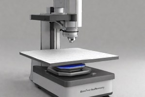
- Instrumentation
- Results and Deliverables
Microelectronics X-Ray Imaging – Overview
Microelectronics X-Ray Imaging is a non-destructive evaluation technique used to inspect internal structures of electronic components and assemblies. It enables visualization of hidden defects such as voids, cracks, misalignments, and solder joint failures without disassembling the device, ensuring accurate failure detection.
The method uses high-energy X-rays to penetrate materials and generate contrast based on density variations. Advanced imaging systems provide high-resolution 2D and 3D views, allowing detailed analysis of internal features. This technique is critical for root cause failure analysis, quality assurance, and reliability assessment in complex microelectronic systems.

Scope, Applications, and Benefits
Scope
Microelectronics X-Ray Imaging focuses on internal inspection of electronic components to detect structural defects and analyze failure mechanisms. It supports non-destructive evaluation and detailed visualization of internal assemblies under controlled conditions.
- Detection of internal defects in electronic components
- Inspection of solder joints and interconnections
- Analysis of voids, cracks, and delamination
- Evaluation of assembly alignment and integrity
- Support for failure analysis and quality control
Applications
- Failure analysis of electronic components
- Inspection of printed circuit board assemblies (PCBs)
- Quality control in electronics manufacturing
- Verification of solder joint integrity
- Detection of hidden defects in semiconductor devices
- Research and development of electronic products
Benefits
- Non-destructive inspection method
- High-resolution internal visualization
- Early detection of hidden defects
- Supports root cause failure analysis
- Improves product reliability and quality
- Reduces need for destructive testing
Microelectronics X-Ray Imaging – Test Process
Sample Positioning
The electronic component is precisely positioned within the X-ray system for optimal imaging.
1X-Ray Exposure
Controlled X-ray beams are passed through the sample to capture internal structural variations.
2Image Acquisition
Detectors capture transmitted X-rays to generate high-resolution images.
3Analysis & Interpretation
Images are analyzed to identify defects and determine root causes of failure.
4Microelectronics X-Ray Imaging – Technical Specification
| Parameter | Details |
|---|---|
| X-Ray Source Type | Microfocus or nanofocus X-ray tube for high-resolution imaging |
| Voltage Range | Typically 20 kV to 160 kV depending on material density and thickness |
| Resolution | Up to 1 µm or better for fine defect detection Detector Type: Flat panel digital detector with high contrast se |
| Magnification | Variable geometric magnification up to 1000× |
| Sample Size | From microchips to large PCB assemblies |
| Imaging Mode | 2D real-time radiography and 3D computed tomography (CT) |
| Scan Time | Seconds for 2D imaging; minutes to hours for 3D CT scans |
| Measurement Output | Void %, crack length, defect size, and positional analysis |
Instrumentation Used for Testing
- X-ray inspection system
- Microfocus X-ray tube
- Flat panel detector
- Computed tomography (CT) system
- Image processing software
- Precision sample stage
- Radiation shielding enclosure
Results and Deliverables
- High-resolution X-ray images (2D/3D)
- Identification of internal defects (voids, cracks, misalignment)
- Quantitative defect measurements
- Failure analysis report with root cause insights
- Comparative analysis with design specifications
- Recommendations for corrective actions
Partnering with Infinita Lab for Optimal Results
Infinita Lab addresses the most frustrating pain points in the microelectronics X-ray imaging process: complexity, coordination, and confidentiality. Our platform is built for secure, simplified support, allowing engineering and R&D teams to focus on what matters most: innovation. From kickoff to final report, we orchestrate every detail—fast, seamlessly, and behind the scenes.
Looking for a trusted partner to achieve your research goals? Schedule a meeting with us, send us a request, or call us at (888) 878-3090 to learn more about our services and how we can support you. Request a Quote

Frequently Asked Questions
Microelectronics X-ray imaging is a non-destructive technique used to inspect internal structures of electronic components. It is used to detect hidden defects, analyze failures, and ensure product reliability without damaging or disassembling the device.
It can detect voids, cracks, solder joint defects, misalignments, delamination, and foreign inclusions. These defects are often invisible externally but can significantly impact performance and reliability.
X-rays pass through the component and are absorbed differently based on material density. The transmitted radiation is captured by detectors to form images that reveal internal structural variations and defects.
Industries such as electronics manufacturing, aerospace, automotive, and semiconductor production benefit from this technique for ensuring reliability and quality.
While it significantly reduces the need for destructive testing, it may be complemented by destructive methods for detailed material analysis when required.


Request a Quote
Submit your material details and receive testing procedures, pricing, and turnaround time within 24 hours.
 Quick Turnaround and Hasslefree process
 Confidentiality Guarantee
 Free, No-obligation Consultation
 100% Customer Satisfaction
