Dual Beam – Focused Ion Beam (Dual Beam – FIB)
Dual Beam - Focused Ion Beam (Dual Beam - FIB) uses a focused ion beam (usually Gallium) for nano-scale milling or sputtering of samples with simultaneous SEM imaging of the modified area. The Dual Beam-FIB technique has many applications in emerging semiconductor and nano-technology sectors and all the tests related to this technique are offered by Infinita Lab network of testing labs, USA, to our clients.

TRUSTED BY


Precision-driven testing for dimensional accuracy and compliance
- Overview
- Scope, Applications, and Benefits
- Test Process
- Specifications
- Instrumentation
- Results and Deliverables
Overview
Dual Beam – Focused Ion Beam (FIB) is a sophisticated microscopy technique that incorporates a focused ion beam and a scanning electron microscope (SEM) in a single tool. With this tool, precise material removal and cross-sectioning of micro and nanostructures are possible.
Dual beam Focused Ion Beam tools are utilized in various applications, ranging from materials and semiconductor analysis to nanotechnology and failure analysis. The tool is utilized to mill or modify a material’s surface with an ion beam and to provide detailed imaging of structures with an electron beam.

Scope, Applications, and Benefits
Scope
Dual Beam FIB analysis enables precise material removal, cross-section preparation, and nanoscale imaging for detailed structural investigation. The technique is particularly useful for examining internal structures and performing site-specific sample preparation.
The scope includes:
Micro and nanoscale material modification
Cross-section preparation of samples
Imaging of internal microstructures
Analysis of semiconductor and electronic devices
Preparation of TEM lamella samples
Applications
Semiconductor device failure analysis
Nanotechnology and microelectronics research
Materials science investigations
Microstructure and interface analysis
TEM sample preparation
Benefits
Enables precise nanoscale milling
Provides high-resolution imaging
Allows site-specific sample preparation
Combines imaging and machining in one system
Supports advanced materials characterization
Test Process
Sample Preparation
The specimen is mounted and positioned in the dual beam chamber for analysis.
1Ion Beam Milling
A focused ion beam removes material from the sample to expose internal structures or prepare cross-sections.
2Electron Beam Imaging
The scanning electron beam captures high-resolution images of the exposed surfaces.
3Data Analysis
The images and structural information are analyzed to evaluate material properties or detect defects.
4Technical Specifications
| Parameter | Details |
|---|---|
| Technique Type | Dual beam microscopy |
| Ion Source | Gallium ion beam |
| Imaging Method | Scanning electron microscopy |
| Resolution | Nanometer-scale imaging capability |
| Operation Mode | Milling, imaging, and sample preparation |
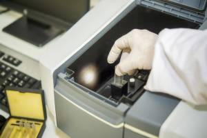
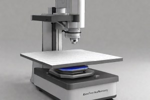
Instrumentation Used for Testing
Dual Beam FIB-SEM System
Focused Ion Beam Source
Scanning Electron Microscope (SEM)
Sample Stage and Chamber
Image Processing Software
Results and Deliverables
High-resolution microstructure images
Cross-sectional analysis reports
Defect and failure analysis results
Nanoscale structural characterization
Detailed technical analysis repor
Why Choose Infinita Lab for Dual Beam – FIB?
With Infinita Lab (www.infinitalab.com), you are guaranteed a Nationwide Network of Accredited Laboratories spread across the USA, the best Consultants from around the world, Convenient Sample Pick-Up and Delivery, and Fast Turnaround Time.
Our team understands the stakes and subtleties of every test. Whether you’re validating a new Product, de-risking a prototype, or navigating complex compliance requirements, our specialists guide the process with rigor and clarity.
Looking for a trusted partner to achieve your research goals? Schedule a meeting with us, send us a request, or call us at (888) 878-3090 to learn more about our services and how we can support you. Request a Quote
Frequently Asked Questions
Dual Beam FIB is an advanced microscopy technique that combines a focused ion beam for precise material removal with a scanning electron microscope for high-resolution imaging and nanoscale structural analysis.
The focused ion beam is used to mill, cut, or remove material from a sample surface, enabling researchers to expose internal structures and prepare precise cross-sections for analysis.
Unlike a conventional SEM, Dual Beam FIB systems include both an ion beam and an electron beam, allowing simultaneous material modification and high-resolution imaging within the same instrument.
Dual Beam FIB enables precise cross-sectioning of microelectronic devices, allowing engineers to examine internal layers, detect defects, and investigate failures in semiconductor components.
Dual Beam FIB systems provide nanoscale resolution imaging and precise material removal, enabling researchers to study microstructures and defects at extremely small dimensions.
Case Studies
In-depth examination of genuine material testing solutions
Dopant and Ultra-Low Concentration Elemental Analysis Using STEM…
Dopant and Ultra-Low Concentration Elemental Analysis Using STEM…
Introduction to STEM-EELS for Elemental Analysis Scanning Transmission Electron Microscopy (STEM) combined with Electron Energy Loss...
Read Case StudyAnalysis of PVC Pipe Degradation Using FTIR Spectroscopy

Analysis of PVC Pipe Degradation Using FTIR Spectroscopy
PVC Pipe in Infrastructure — and Why Degradation Matters Polyvinyl chloride (PVC) pressure pipe is one...
Read Case StudyNano-scale roughness measurement of Si-wafers by Atomic Force…
Nano-scale roughness measurement of Si-wafers by Atomic Force…
Nano-scale surface roughness is a critical parameter in fabricated thin-films that are used in optics, solar...
Read Case Study
Request a Quote
Submit your material details and receive testing procedures, pricing, and turnaround time within 24 hours.
 Quick Turnaround and Hasslefree process
 Confidentiality Guarantee
 Free, No-obligation Consultation
 100% Customer Satisfaction
