Digital Radiography
Digital radiography is non-destructive technique frequently used for material inspection in the aerospace, automotive, and industrial manufacturing sectors.

TRUSTED BY


Precision-driven testing for dimensional accuracy and compliance
- Overview
- Scope, Applications, and Benefits
- Test Process
- Specifications
- Instrumentation
- Results and Deliverables
Overview
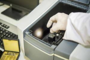
Digital Radiography (DR) is a non-destructive imaging technique that uses X-rays and advanced digital detectors to capture high-resolution images of internal structures without the need for film processing. It enables real-time visualization, faster inspection cycles, and improved accuracy in detecting internal defects.
This technique is widely used in the industrial inspection, manufacturing, and materials testing industry for evaluating structural integrity, weld quality, and component reliability. Its ability to provide instant digital results, enhanced image processing, and easy data storage makes it a preferred method for modern quality assurance and failure analysis applications.

Scope, Applications, and Benefits
Scope
Digital radiography enables fast and accurate internal inspection of components, identifying defects and structural inconsistencies without damaging the sample.
It supports quality assurance, failure analysis, and inspection of complex assemblies across various materials.
- Internal defect detection (cracks, voids, inclusions)
- Weld inspection and evaluation
- Casting and component inspection
- Thickness and density variation analysis
- Assembly verification
- Non-destructive testing (NDT)
- Real-time imaging and analysis
Applications
- Welded joints and pipelines
- Castings and metal components
- Aerospace and automotive parts
- Electronic assemblies and PCBs
- Composite materials
Benefits
- Non-destructive inspection method
- Immediate digital image results
- High-resolution defect detection
- Reduced inspection time
- Easy data storage and sharing
- Improved accuracy and reliability
- Cost-effective for repeated inspections
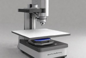
Test Process
Sample Positioning
Component is positioned between the X-ray source and digital detector.
1Exposure Setup
X-ray parameters like voltage and exposure time are adjusted.
2Image Acquisition
X-rays pass through the sample and are captured by the detector.
3Image Analysis
Digital images are analyzed to identify internal defects or variations.
4Technical Specifications
| Parameter | Details |
|---|---|
| Material Type | Metals, composites, plastics, and electronic assemblies. |
| X-ray Voltage Range | Typically 40 kV to 450 kV depending on material thickness. |
| Detector Type | Flat panel digital detectors or computed radiography plates. |
| Image Resolution | High-resolution imaging for defect detection. |
| Exposure Time | Seconds to minutes based on material and thickness. |
| Image Format | Digital images stored in standard formats. |
| Software Analysis | Image processing and defect evaluation tools. |
Instrumentation Used for Testing
- X-ray generator
- Digital flat panel detector
- Computed radiography system
- Image processing software
- Positioning fixtures
- Radiation shielding equipment
- Monitoring devices
Results and Deliverables
- Digital radiographic images
- Defect identification reports
- Weld quality evaluation
- Thickness and density analysis
- Inspection documentation
- Test reports (ASTM/ISO)
- Final test certification
Partnering with Infinita Lab for Optimal Results
Infinita Lab addresses the most frustrating pain points in the digital radiography testing process: complexity, coordination, and confidentiality. Our platform is built for secure, simplified support, allowing engineering and R&D teams to focus on what matters most: innovation. From kickoff to final report, we orchestrate every detail—fast, seamlessly, and behind the scenes.
Looking for a trusted partner to achieve your research goals? Schedule a meeting with us, send us a request, or call us at (888) 878-3090 to learn more about our services and how we can support you. Request a Quote
Frequently Asked Questions
Digital radiography is a non-destructive testing method that uses X-rays and digital detectors to inspect internal structures of materials, helping identify defects without damaging the component.
X-rays pass through a material and are captured by a digital detector, producing an image that reveals internal features such as cracks, voids, and structural inconsistencies.
It provides instant results, high-resolution images, easy data storage, and improved inspection efficiency compared to traditional film-based radiography methods.
DR uses digital detectors for instant image capture, while conventional radiography uses film, requiring processing time and additional steps.
Cracks, voids, inclusions, porosity, and weld defects can be identified using digital radiography.
Case Studies
In-depth examination of genuine material testing solutions
Dopant and Ultra-Low Concentration Elemental Analysis Using STEM…
Dopant and Ultra-Low Concentration Elemental Analysis Using STEM…
Introduction to STEM-EELS for Elemental Analysis Scanning Transmission Electron Microscopy (STEM) combined with Electron Energy Loss...
Read Case StudyAnalysis of PVC Pipe Degradation Using FTIR Spectroscopy

Analysis of PVC Pipe Degradation Using FTIR Spectroscopy
PVC Pipe in Infrastructure — and Why Degradation Matters Polyvinyl chloride (PVC) pressure pipe is one...
Read Case StudyNano-scale roughness measurement of Si-wafers by Atomic Force…
Nano-scale roughness measurement of Si-wafers by Atomic Force…
Nano-scale surface roughness is a critical parameter in fabricated thin-films that are used in optics, solar...
Read Case Study
Request a Quote
Submit your material details and receive testing procedures, pricing, and turnaround time within 24 hours.
 Quick Turnaround and Hasslefree process
 Confidentiality Guarantee
 Free, No-obligation Consultation
 100% Customer Satisfaction
