


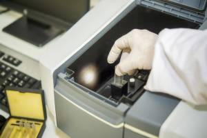
ASTM E1165 X-Ray Focal Spot Measurement
Focal spots of industrial X-Ray tubes are measured by pinhole imaging, using ASTM E1165 standard test method. This test method specifies the process to determine the size of standard and mini focal spots in industrial X-ray tubes with focal spot dimensions ranging from 100 m to several mm and a tube voltage of 600 kV. Values expressed in SI units are considered standard.

TRUSTED BY


ASTM E1165 X-Ray Focal Spot Measurement
- Overview
- Scope, Applications, and Benefits
- Test Process
- Specifications
- Instrumentation
- Results and Deliverables
ASTM E1165 X-Ray Focal Spot Measurement Overview
ASTM E1165 is the standard test method for measuring the size and intensity distribution of focal spots in industrial X-ray tubes using the pinhole camera technique. The focal spot is the area on the X-ray tube anode where the electron beam strikes and X-rays are produced. Its size directly determines the geometric unsharpness of radiographic images -a smaller focal spot produces sharper images that resolve finer detail. In comparison, a larger focal spot creates penumbral blur that limits the minimum detectable flaw size in radiographic inspection.
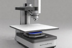
The pinhole imaging method works by placing a small aperture -the pinhole -between the X-ray tube and a recording medium such as film or a digital detector. The pinhole projects a magnified image of the focal spot onto the detector, and the resulting image is measured to determine the focal spot dimensions in two orthogonal directions: width (perpendicular to the tube axis) and length (along the tube axis). The magnification factor, determined by the geometry of the pinhole setup, is used to calculate the actual focal spot dimensions from the image measurements.
Accurate focal spot measurement matters for several reasons. It is required when qualifying X-ray equipment for use on radiographic procedures that specify maximum allowable geometric unsharpness. It is also used during tube acceptance, periodic verification, and when a tube is replaced or repaired to confirm that it meets the manufacturer’s claimed focal spot size. The standard covers focal spot dimensions from 100 micrometers up to several millimeters and tube voltages up to 600 kV.

ASTM E1165 X-Ray Focal Spot Measurement Scope, Applications, and Benefits
Scope
ASTM E1165 covers the measurement of focal spot size and intensity distribution for standard and mini focal spots in industrial X-ray tubes using the pinhole camera method. The standard applies to focal spot dimensions from 100 micrometers to several millimeters and tube voltages up to 600 kV. The pinhole aperture is positioned between the X-ray source and a recording medium at defined source-to-pinhole and pinhole-to-detector distances to produce a magnified focal-spot image. Measurements are made in both the width and length directions of the focal spot image. The standard defines criteria for selecting pinhole size based on nominal focal spot size, magnification factor requirements, and image recording and measurement procedures. Results are reported in SI units (millimeters). The standard falls under the jurisdiction of ASTM Committee E07 on Nondestructive Testing.
Applications
- X-ray tube acceptance testing to verify focal spot size against manufacturer specifications
- Periodic focal spot verification for X-ray systems used in production radiographic inspection
- Equipment qualification for radiographic procedures specifying maximum geometric unsharpness limits
- Focal spot documentation for radiographic procedure qualification records
- Post-repair or post-replacement tube verification following anode or tube head service
- Microfocus and minifocus X-ray tube characterization for high-resolution radiographic inspection
- Computed tomography (CT) system focal spot verification affecting reconstruction resolution
- Comparison of focal spot size across operating conditions -different kV settings, mA levels, or focal spot modes
Benefits
- Provides a direct physical measurement of the focal spot rather than relying on manufacturer data alone
- Pinhole method is traceable and reproducible -the same geometry yields comparable results across labs and over time.
- Covers the full range of industrial X-ray tube focal spot sizes from microfocus to standard large focal spots
- Results feed directly into geometric unsharpness calculations used in radiographic procedure qualification.n
- Required documentation for many aerospace, nuclear, and pressure vessel radiographic procedures
- Identifies focal spot changes due to tube aging, anode degradation, or operating condition shifts
- Applicable to both film-based and digital radiographic recording media
ASTM E1165 X-Ray Focal Spot Measurement Process
Setup and Geometry Establishment
The pinhole aperture is selected based on the nominal focal spot size per the standard's selection criteria.
1X-Ray Exposure
The X-ray tube is operated at the specified technique factors -tube voltage (kV) and tube current (mA) appropriate to the focal spot size being measured.
2Image Processing and Measurement
The recorded image is processed, and the focal spot profile is extracted.
3Reporting
Focal spot width and length are reported in millimeters at the specified measurement threshold.
4ASTM E1165 X-Ray Focal Spot Measurement Technical Specifications
| Parameter | Details |
|---|---|
| Standard | ASTM E1165-20 |
| ASTM Committee | E07 – Nondestructive Testing |
| Measurement Method | Pinhole camera imaging |
| Focal Spot Size Range | 100 micrometers to several millimeters |
| Tube Voltage Range | Up to 600 kV |
| Measurement Directions | Width (perpendicular to tube axis) and length (along tube axis) |


Instrumentation Used for ASTM E1165 X-Ray Focal Spot Measurement
- Calibrated pinhole aperture of appropriate diameter for the focal spot size being measured
- Pinhole holder with precise positioning and alignment capability
- Industrial X-ray film or digital flat panel detector for image recording
- Densitometer for film-based image analysis
- Digital image analysis software for focal spot profile measurement and dimension extraction
- Precision ruler or calibrated scale for geometry verification and magnification factor confirmation
- Alignment tools for centering the pinhole on the central beam axis
ASTM E1165 X-Ray Focal Spot Measurement Results and Deliverables
- Measured focal spot width in millimeters
- Measured focal spot length in millimeters
- Focal spot intensity distribution profile image in both measurement directions
- Magnification factor used and source-to-pinhole and pinhole-to-detector distances
- Tube operating conditions at time of measurement -kV, mA, focal spot mode
- Pinhole aperture diameter and material
- Comparison of measured focal spot dimensions against nominal or specified maximum size
- Recording medium type and processing details
- Test report with all measured values, images, geometry documentation, and equipment identification formatted for equipment qualification records or procedure documentation
Frequently Asked Questions
Results can be influenced by porosity, firing temperature, microstructural defects, specimen geometry, surface finish, and internal residual stresses from processing.
A ceramic specimen is subjected to a controlled bending load until fracture occurs, typically using a three-point or four-point bending setup. The breaking stress is then calculated.
ASTM C674 determines the flexural strength (modulus of rupture) of ceramic whiteware under bending load. It helps assess how the material behaves under mechanical stress before fracture.
Flexural strength indicates resistance to cracking or breaking during handling, processing, and end-use. It is a key quality parameter for structural reliability in ceramic products.
The method is used for ceramic whiteware such as porcelain, stoneware, sanitary ware, and electrical ceramics where brittle fracture behavior must be characterized.
Why Choose Infinita Lab for Advanced Materials Testing and Characterization?
At the core of this breadth is our network of 2,000+ accredited laboratories across the USA, offering access to over 10,000 testing methods and analytical services. From advanced materials characterization (SEM, TEM, RBS, XPS) to mechanical, chemical, environmental, biological, and standardized ASTM/ISO-compliant testing, we deliver unmatched flexibility, specialization, and scale. You are never limited by geography, facility, or methodology — Infinita Lab connects you to the right expertise and testing solution, every time.

Looking for a Trusted Partner for Accurate and Reliable Testing Services?
Send query us at hello@infinitlab.com or call us at (888) 878-3090 to learn more about our services and how we can support you.

Request a Quote
Submit your material details and receive testing procedures, pricing, and turnaround time within 24 hours.
 Quick Turnaround and Hasslefree process
 Confidentiality Guarantee
 Free, No-obligation Consultation
 100% Customer Satisfaction
