X-Ray Fluorescence (XRF) and its Applications
X-Ray Fluorescence (XRF) has a variety of applications in different fields. Learn more about its applications and its basics.
A Definition of X-Ray Fluorescence (XRF)
Regular, disruptive chemical examinations of rocks, minerals, sediments, and fluids are performed with the help of an X-ray fluorescence (XRF) spectrometer. It uses the same sort of wavelength-dispersive spectroscopy as an electron microprobe (EPMA) to do its work. However, an XRF is often employed for bulk investigations of larger fractions of geological materials, and it cannot generally do analyses at the small spot sizes characteristic of EPMA work (2-5 microns). X-ray spectroscopy is one of the most popular methods for analyzing major and trace elements in rocks, minerals, and sediment because of its stability, low cost, and convenience of use.
Fluorescence X-Ray Analysis: The Basics
X-ray fluorescence (XRF) relies on the same fundamental principles as X-ray spectroscopy (e.g., scanning electron microscopy – energy dispersive spectroscopy), X-ray diffraction (XRD), and wavelength dispersive spectroscopy (microprobe WDS).
Atomic behaviour upon interaction with radiation allows for investigating main and trace elements in geological materials using X-ray fluorescence. Ionization occurs in materials when they are stimulated by high-energy, short-wavelength radiation (like X-rays). Suppose the radiation is powerful enough to knock a closely bound inner electron loose. In that case, the atom will become unstable, and another electron from its outer shell will move in and take its place. Since the binding energy of an inner electron orbital is less than that of an outer one, energy is liberated in this process. The released radiation, known as fluorescence radiation, has less energy than the main incident X-rays. The resultant fluorescence X-rays can be utilized to determine the abundances of elements in the sample due to the energy of the emitted photon indicative of a transition between specific electron orbitals in a particular element.
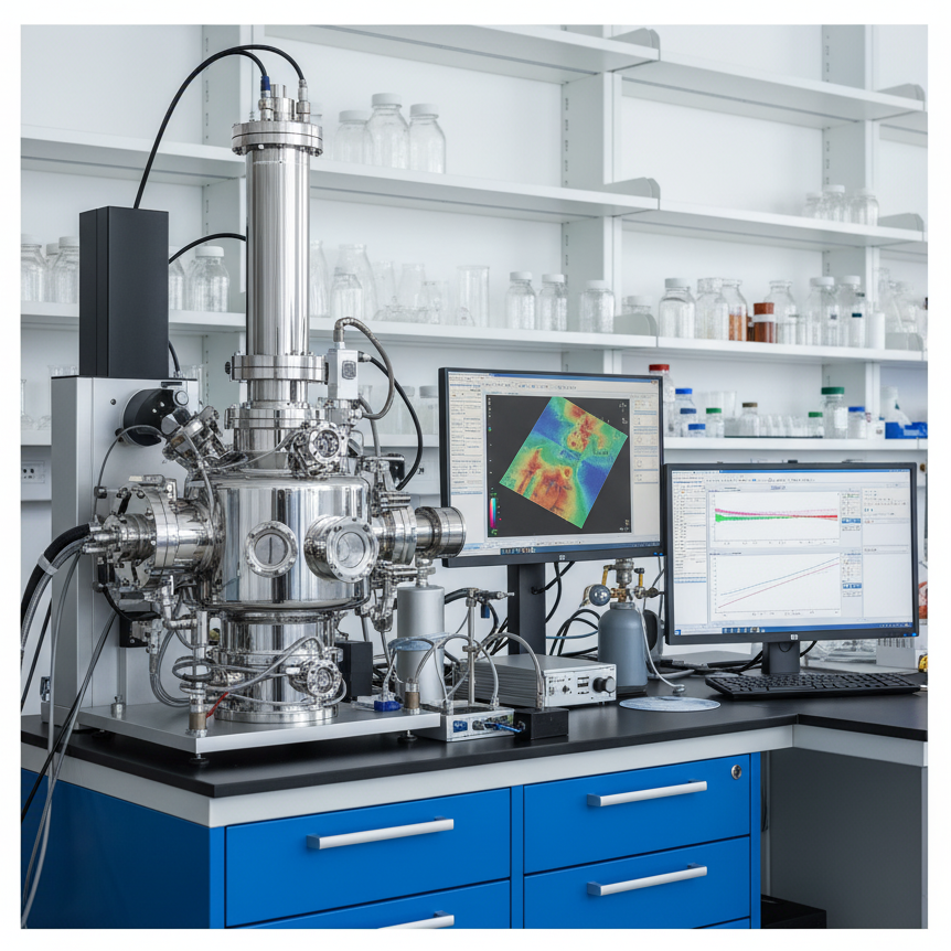
Fluorescence X-Ray Spectrometry (XRF) Equipment – How Does It Operate?
Geological material analysis through X-ray fluorescence (XRF) relies on the interaction between atoms and X-rays to enable the detection of major and trace elements. A sample is subjected to an intense X-ray beam called the incident beam. Depending on the sample’s chemical makeup, some of the energy is absorbed by the sample itself, and some is scattered. While Rh targets are frequently used, other materials like W, Mo, and Cr can also produce the incident X-ray beam.
Sample stimulation occurs when the sample is exposed to this initial X-ray beam. The stimulated sample reacts by releasing X-rays with a wavelength spectrum determined by the sorts of atoms it contains. Why does this happen? Ionisation is the process by which the sample’s atoms absorb X-ray radiation and liberate electrons from their lower energy states (often K and L). The released electrons are replaced by electrons from a higher-energy, farther-off orbit. Energy is released during this process because the binding energy of an inner electron orbital is lower than that of an outer one. It is possible to determine the kind of atom emitting by analysing the released X-rays. Most minerals and rocks include many elements, which necessitates the employment of a Wavelength Dispersive Spectrometer similar to that found in an EPMA in order to disentangle the complex emitted X-ray spectrum into the characteristic wavelengths for each of those elements. Among other instruments, the beam’s strength is measured by scintillation and gas flow proportional detectors. The K spectra of elements lighter than zinc, which usually generate X-rays with a large wavelength (>0.15 nm), are commonly measured using the flow counter. A scintillation detector is typically used to analyse the X-ray spectrum at shorter wavelengths (K spectra of elements from Nb through I; L spectra of Th and U). Both detectors are usually used in conjunction when measuring intermediate-wavelength X-rays. These detectors measure the energy present in a sample in a manner directly proportional to the element’s abundance. The precise value of this proportionality for each element is determined by comparing the sample to known compositional mineral or rock standards.
X-Ray Fluorescence (XRF) Applications
- X-ray fluorescence can benefit soil surveys, investigations of igneous, sedimentary, and metamorphic petrology, and much more.
- Quality control is essential in many industries, including mining (for example, for determining ore grade), cement production, ceramic and glass manufacture, and metallurgy.
- Studies of the natural world (such as the composition of dust on air filters)
- Sulfur levels in crude oils and finished petroleum products are a major concern for the petroleum industry.
- Portable X-ray fluorescence spectrometers (for use in geological and environmental research) are also used in the field.
- Bulk chemical analyses of major elements (Si, Ti, Al, Fe, Mn, Mg, Ca, Na, K, P) and trace elements (in abundances >1 ppm; Ba, Ce, Co, Cr, Cu, Ga, La, Nb, Ni, Rb, Sc, Sr, Rh, U, V, Y, Zr, Zn) in rock and sediment are ideal applications for X-ray fluorescence. – The lowest detectable concentrations of trace elements are on the order of a few ppm.
- X-ray fluorescence analysis is restricted to large samples (typically > 1 gram) that can be prepared as a powder and homogenized effectively; materials for which compositionally similar, well-characterized standards are available; materials containing high abundances of elements for which absorption and fluorescence effects are reasonably well understood; and materials for which X-rays can be efficiently absorbed by the sample.
- When working with rocks, ores, sediments, and minerals, grinding the material to a powder is standard practice. This allows for direct analysis, which is useful for analyzing trace elements. The proportionality comparison to the standards is complicated by the vast variation in abundances of several elements, especially iron, and the diversity in particle sizes present in a powdered sample. This is why it is standard practice to combine the powdered sample with a chemical flux before melting it in a furnace or gas burner. The abundances of the (now somewhat diluted) elements can be estimated by melting the material into a homogeneous glass, which can then be examined.
Where Does X-Ray Fluorescence (XRF) Shine, and Where Does It Fall Short?
Strengths
Particular fields of study that benefit greatly from X-ray fluorescence analysis are:
- Analysis of rocks and sediments for their principal chemical components (Si, Ti, Al, Fe, Mn, Mg, Ca, Na, K, and P)
- Rock and sediment samples were analyzed chemically for the presence of trace elements (>1 ppm; Ba, Ce, Co, Cr, Cu, Ga, La, Nb, Ni, Rb, Sc, Sr, Rh, U, V, Y, Zn).
X-Ray Fluorescence (XRF) Test Limitations
- Depending on the wavelength and intensity of the input X-rays, the XRF can theoretically detect X-ray emissions from nearly all elements.
- The capacity of commonly accessible commercial tools to precisely and correctly determine the abundance of elements with Z11 in most natural earth materials is severely lacking.
- Different isotopes of an element can be analyzed with different equipment (see TIMS and SIMS), as XRF analyses cannot do so.
- Since wet chemical analysis and Mossbauer spectroscopy can distinguish between ions of the same element in different valence states, these methods are used instead of XRF examinations when analyzing rocks and minerals.
Instructions for Taking and Preparing Samples
The availability of suitable standards allows the analysis of nearly any solid or liquid. The usual commercial instrument requires a sample of at least several grams of material for rocks and minerals, though the gathered sample may be much greater. In order to conduct an accurate XRF chemical analysis of a rock, a sample larger than the largest grain or particle in the rock must be gathered. After undergoing a series of crushing stages, the initial sample is reduced to an average grain size of a few millimetres to a centimetre. At this point, it can be further divided to yield a tiny representative sample weighing in the tens to hundreds of grams. The XRF sample is created by grinding a small sample and splitting it into a fine powder using several methods. At this stage, it is especially important to consider the possibility of contamination due to the crushing tools’ material.
Collecting Information, Analyzing It, and Reporting It
- Spectrum of X-Rays
- Limits of detection data table
- Punctuality and Accuracy
- Quality Assessment of Data (flyers, trends, discriminant fields) using Database and Plotting
- Chemical Maps
 Metrology Testing Service

- Gas Chromatography (GC) Analysis Services
- Nuclear Magnetic Resonance (NMR) Spectroscopy Testing Services
- Plasma Focused Ion Beam (P-FIB): How It Works & Applications
- Electronic Failure Analysis Using Scanning Acoustic Microscopy (SAM)
- Current Leakage Detection: Tools, Techniques & Electrical Testing Standards
- Powder X-Ray Diffraction (PXRD): Crystalline Material Analysis & Phase Identification
- Transmission Electron Microscopy (TEM) Analysis Services
- Semiconductor Failure Analysis Techniques: A Complete Overview
- View All


3 Easy Steps to Start Testing


Case Studies
In-depth examination of genuine material testing solutions
Case Study: Dopant & Ultra-Low Concentration Analysis via…

Case Study: Dopant & Ultra-Low Concentration Analysis via…
Introduction to STEM-EELS for Elemental Analysis Scanning Transmission Electron Microscopy (STEM) combined with Electron Energy Loss...
Read Case StudyAFM Nano-Scale Roughness Measurement Guide for Silicon Wafers
AFM Nano-Scale Roughness Measurement Guide for Silicon Wafers
Nano-scale surface roughness is a critical parameter in fabricated thin-films that are used in optics, solar...
Read Case StudySolid-state Li batteries failure analysis using Scanning Electron…

Solid-state Li batteries failure analysis using Scanning Electron…
SEM imaging is used to understand the effects of gold thin-films as an interface modifier to...
Read Case StudyTalk to Our Experts Today!
Submit your contact info and we’ll get back to you within 24 hours
