


Photo-Induced Force Microscopy (PiFM) – Nanoscale IR Chemical Imaging
Photo-induced force microscopy combines Atomic Force Microscopy (AFM) and Infra-red (IR) spectroscopy principles, to perform chemical mapping of photo-polarizable molecular species, at nano-scale. It is applicable to both organic and inorganic molecules. In this case study, a polymer blend sample of Poly Lactic acid (PLA), Acrylic rubber (ACM) and a third component, was analysed by PiFM to identify and map polymer distribution across the surface.

TRUSTED BY


Precision Testing Solutions for Accurate, Reliable, and Standards-Compliant Results
- Overview
- Scope, Applications, and Benefits
- Test Process
- Specifications
- Instrumentation
- Results and Deliverables
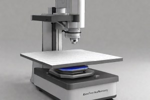
Photo Induced Force Microscopy (PIFM) – Overview
Photo Induced Force Microscopy (PiFM) is an advanced nanoscale characterization technique that combines atomic force microscopy with infrared spectroscopy to map chemical composition with high spatial resolution. It detects photo-induced dipole–dipole interactions between a sample and a probe under localized optical excitation.
PiFM enables simultaneous topographical and chemical imaging beyond the diffraction limit of light. By measuring forces generated from light–matter interaction, it provides molecular-level insights into heterogeneous materials, making it highly effective for analyzing polymers, nanomaterials, and thin films.

Scope, Applications, and Benefits
Scope
PIFM focuses on nanoscale chemical characterization by detecting photo-induced forces generated through localized optical excitation and probe interaction.
It includes simultaneous mapping of chemical composition and surface morphology, enabling identification of material distribution, phase separation, and molecular interactions at nanometer-scale resolution.
- Nanoscale chemical imaging of heterogeneous materials
- Simultaneous topography and chemical mapping
- Identification of molecular composition and distribution
- Analysis of polymers, nanomaterials, and thin films
- Detection of phase separation and domain structures
- Evaluation of local chemical heterogeneity
- High-resolution spectroscopy beyond diffraction limits
- Correlation of physical and chemical properties
Applications
- Polymer and composite analysis
- Nanotechnology and material science research
- Thin film characterization
- Semiconductor material analysis
- Surface chemistry studies
- Advanced coating evaluation
Benefits
- Provides nanoscale chemical resolution
- Combines imaging and spectroscopy in one technique
- Enables analysis beyond optical diffraction limits
- Offers high sensitivity to molecular interactions
- Supports detailed material characterization
- Enhances understanding of nanoscale structures
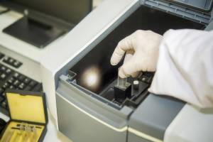
Photo Induced Force Microscopy (PIFM) – Process
Sample Preparation & Surface Conditioning Procedure
Samples are prepared with smooth, clean surfaces to ensure accurate probe interaction and signal detection.
1AFM Probe Alignment & Optical Excitation Setup
A sharp probe is aligned while a tunable infrared laser excites the sample at specific wavelengths.
2Photo-Induced Force Detection & Signal Acquisition Stage
Forces generated by dipole interactions are measured as the probe scans the surface.
3Spectral Analysis & Nanoscale Chemical Mapping Reporting
Data is processed to generate chemical maps and spectra with nanometer-scale resolution.
4Photo Induced Force Microscopy (PiFM) – Technical Specification
| Parameter | Details |
|---|---|
| Test Method | AFM-based photo-induced force detection with infrared excitation |
| Measurement Type | Nanoscale chemical and topographical mapping |
| Sample Type | Polymers, thin films, nanomaterials, and composites |
| Loading Type | Probe-sample interaction under optical excitation |
| Units | Force (pN), wavelength (nm or cm⁻¹), spatial resolution (nm) |
| Spatial Resolution | Typically below 10 nm |


Instrumentation Used for Testing
- Atomic force microscope
- Tunable infrared laser source
- PiFM detection module
- Vibration isolation system
- Optical alignment system
- Data acquisition and analysis software
Results and Deliverables
- Nanoscale chemical maps
- Infrared spectra of localized regions
- Surface topography images
- Phase and composition distribution data
- Material heterogeneity analysis
- Detailed analytical report
Frequently Asked Questions
PiFM measures localized forces generated by light–matter interaction at the probe tip rather than relying on far-field optics, enabling spatial resolution determined by tip size rather than wavelength.
The technique detects dipole–dipole interactions created when the sample absorbs infrared light, generating localized forces that are measured by the AFM probe.
Conventional spectroscopy provides bulk information, while PiFM offers nanoscale chemical mapping by combining spectroscopy with atomic force microscopy.
By tuning the excitation wavelength to specific vibrational resonances, PiFM selectively enhances contrast between chemically similar domains, enabling distinction based on subtle differences in molecular bonding and absorption characteristics at nanometer scales.
Variations in tip geometry, local field enhancement, and sample optical properties can influence signal intensity, making absolute quantification challenging without careful calibration and controlled experimental conditions.
Why Choose Infinita Lab for Advanced Materials Testing and Characterization?
At the core of this breadth is our network of 2,000+ accredited laboratories across the USA, offering access to over 10,000 testing methods and analytical services. From advanced materials characterization (SEM, TEM, RBS, XPS) to mechanical, chemical, environmental, biological, and standardized ASTM/ISO-compliant testing, we deliver unmatched flexibility, specialization, and scale. You are never limited by geography, facility, or methodology — Infinita Lab connects you to the right expertise and testing solution, every time.

Looking for a Trusted Partner for Accurate and Reliable Testing Services?
Send query us at hello@infinitlab.com or call us at (888) 878-3090 to learn more about our services and how we can support you.
Request a Quote
Submit your material details and receive testing procedures, pricing, and turnaround time within 24 hours.
 Quick Turnaround and Hasslefree process
 Confidentiality Guarantee
 Free, No-obligation Consultation
 100% Customer Satisfaction
