


Fluorescent Microthermal Imaging (FMI) Testing for IC Hot Spot Detection
A method for observing and analyzing temperature distribution on a small scale is fluorescent microthermal imaging. It combines thermal imaging with fluorescence microscopy, in which a sample is treated with a temperature-sensitive fluorescent dye. Researchers may detect and measure temperature swings with great spatial resolution because of the dye's temperature-dependent fluorescence emission. Materials science, biology, and other disciplines where precise microscale temperature mapping is necessary can all benefit from this method.

TRUSTED BY


Precision-driven testing for dimensional accuracy and compliance
- Overview
- Scope, Applications, and Benefits
- Test Process
- Specifications
- Instrumentation
- Results and Deliverables
Overview
Fluorescent Microthermal Imaging (FMI) is a high-resolution thermal mapping technique used in electronics failure analysis and reliability testing. It detects temperature variations across integrated circuit surfaces by measuring changes in the fluorescent emission intensity of a temperature-sensitive liquid crystal or fluorescent dye layer applied to the device surface.
FMI provides two-dimensional thermal maps with sub-micron spatial resolution and millikelvin temperature sensitivity, enabling precise localisation of hot spots, resistive faults, thermal runaway sites, and power dissipation anomalies invisible to conventional infrared thermography.

Scope, Applications, and Benefits
Scope
Failure Mode Imaging (FMI) is used to evaluate thermal and electrical behaviour in semiconductor and electronic devices with high spatial precision. It enables micron-scale mapping of surface temperature distribution, helping identify localised thermal anomalies and performance issues during operation.
Key evaluation areas include:
- Surface temperature distribution at micron-scale spatial resolution for detailed thermal mapping
- Hot spot localisation in active IC devices to detect overheating regions
- Resistive and Joule heating fault sites for failure analysis and root cause identification
- Thermal uniformity assessment of power devices and LED arrays to ensure consistent performance
- Temperature coefficient of resistance (TCR) characterisation to study resistance changes with temperature
Applications
- IC hot spot detection and failure analysis
- Power MOSFET and IGBT thermal mapping
- LED junction and thermal resistance characterisation
- PCB trace and via hot spot identification
- MEMS thermal actuator characterisation
Benefits
- Sub-micron spatial resolution (superior to IR microscopy)
- Non-contact, non-destructive measurement
- Millikelvin temperature resolution
- Compatible with packaged and bare die devices
- Enables correlation with emission microscopy findings
Test Process
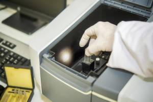
Device Preparation
A temperature-sensitive fluorescent layer is applied and the device is mounted.
1Electrical Biasing
The device is biased under target operating conditions.
2Fluorescence Imaging
UV excitation and CCD imaging are used to generate a temperature map.
3Thermal Analysis
Hot spots and thermal gradients are analyzed for defect localization.
4Technical Specifications
| Parameter | Details |
|---|---|
| Spatial Resolution | 0.3–1 µm (objective dependent) |
| Temperature Sensitivity | 0.1–1 mK |
| Temperature Range | 20–150 °C (standard liquid crystal) |
| Excitation | UV (365 nm typical) |
| Camera | Cooled monochrome CCD |


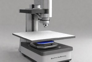
Instrumentation Used for Testing
- Fluorescent microthermal imaging microscope
- Temperature-calibrated fluorescent liquid crystal layer
- UV illumination source
- Cooled CCD camera
- Precision probe station and current source
- Image processing and temperature calibration software
Results and Deliverables
- 2D temperature distribution maps (colour-coded)
- Hot spot coordinates and peak temperature values
- Temperature profiles and cross-section line scans
- Overlay of thermal map with device layout image
- Failure analysis report with thermal findings and defect classification
Frequently Asked Questions
FMI achieves spatial resolution of <1 µm, while conventional IR thermography is limited to ~3–10 µm. FMI is preferred for sub-micron defect localization in advanced IC devices where IR resolution is insufficient.
The applied layer is extremely thin (typically <1 µm) and does not significantly affect thermal or electrical performance. However, it may slightly alter optical reflectance and must be removed before subsequent deprocessing steps.
For packaged devices, top-surface access is required. Packages may need to be opened or decapped to expose the die surface for fluorescent layer application and imaging.
Standard liquid crystal layers operate in the 20–150 °C range. High-temperature formulations are available for devices operating at elevated temperatures, though these may have reduced sensitivity.
FMI is used after electrical characterization to localize thermal anomalies, often in parallel with emission microscopy. Together they guide targeted deprocessing, FIB cross-sectioning, or chemical analysis to identify the physical root cause.
Why Choose Infinita Lab for Advanced Materials Testing and Characterization?
At the core of this breadth is our network of 2,000+ accredited laboratories across the USA, offering access to over 10,000 testing methods and analytical services. From advanced materials characterization (SEM, TEM, RBS, XPS) to mechanical, chemical, environmental, biological, and standardized ASTM/ISO-compliant testing, we deliver unmatched flexibility, specialization, and scale. You are never limited by geography, facility, or methodology — Infinita Lab connects you to the right expertise and testing solution, every time.

Looking for a Trusted Partner for Accurate and Reliable Testing Services?
Send query us at hello@infinitlab.com or call us at (888) 878-3090 to learn more about our services and how we can support you.

Request a Quote
Submit your material details and receive testing procedures, pricing, and turnaround time within 24 hours.
 Quick Turnaround and Hasslefree process
 Confidentiality Guarantee
 Free, No-obligation Consultation
 100% Customer Satisfaction
