Focused Ion Beam SEM (FIB-SEM)
Focused Ion Beam Scanning Electron Microscopy (FIB-SEM) is an advanced analytical technique used in materials science, engineering, and biology to study the microstructure and properties of materials at high resolution. It combines the high-resolution imaging capability of SEM with the precision milling capabilities of a focused ion beam.

TRUSTED BY


Precision-driven testing for dimensional accuracy and compliance
- Overview
- Scope, Applications, and Benefits
- Test Process
- Specifications
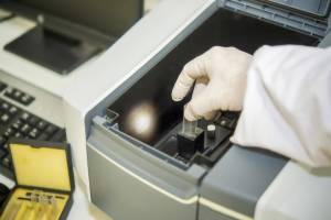
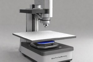
- Instrumentation
- Results and Deliverables
FIB-SEM - Overview
Focused Ion Beam Scanning Electron Microscopy (FIB-SEM) is a dual-beam analytical platform combining a focused gallium ion beam for precision micro/nanoscale material removal with a high-resolution scanning electron microscope for real-time imaging. FIB-SEM enables site-specific cross-sectioning, TEM lamella preparation, 3D tomography, and nanoscale microfabrication on virtually any solid material.
This technique is indispensable in semiconductor failure analysis, materials characterisation, and nanotechnology, where direct access to subsurface features at nanometre resolution is required for root cause analysis, process development, and device characterisation.

Scope, Applications, and Benefits
Scope
Focused Ion Beam–Scanning Electron Microscopy (FIB-SEM) is a powerful technique for high-resolution subsurface analysis and site-specific sample preparation. It enables precise cross-sectioning and imaging, making it ideal for evaluating structural integrity and microstructural features at the nanoscale.
FIB-SEM is used to evaluate:
- Subsurface defects such as voids, delamination, and cracks
- Layer thickness and interface quality in multilayer structures
- Grain structure and morphology at cross-sections
- Site-specific TEM sample preparation (lamella extraction)
- 3D volumetric reconstruction of microstructures for detailed analysis
Applications
- Semiconductor failure analysis (FA) and process control
- TEM sample preparation for advanced node devices
- Corrosion and coating cross-section characterisation
- Battery electrode and separator cross-section analysis
- Metallurgical grain boundary and precipitate study
Benefits
- Site-specific cross-sectioning with <10 nm precision
- Real-time SEM imaging during FIB milling
- Simultaneous EDS/EBSD acquisition on cross-sections
- Enables TEM analysis of targeted subsurface features
- 3D reconstruction reveals volumetric microstructural information
FIB-SEM - Test Process
Site Identification & Protection
Locate ROI and deposit protective layer to prevent beam damage.
1Cross-Section Milling
Use FIB to mill and expose cross-section with high precision.
2Imaging & Analysis
Perform SEM imaging with EDS/EBSD as needed.
3TEM Lamella Prep
Extract and thin lamella (~100 nm) for TEM analysis (if required).
4FIB-SEM - Technical Specifications
| Parameter | Details |
|---|---|
| Ion Beam | Ga⁺ focused ion beam (1–30 kV, 1 pA–65 nA) |
| SEM Resolution | 1–1.5 nm (field emission) |
| Milling Precision | <10 nm positional accuracy |
| Integrated Detectors | SE, BSE, in-lens, EDS, EBSD, STEM |
| Sample Compatibility | All solid materials (conductive and non-conductive) |
Instrumentation Used for Testing
- Dual-beam FIB-SEM system (Thermo Fisher Helios, ZEISS Crossbeam, etc.)
- GIS (gas injection system) for Pt/C deposition
- In-situ micromanipulator (Omniprobe/Kleindiek) for lamella extraction
- EDS and EBSD detectors
- 3D tomography and reconstruction software
Results and Deliverables
- High-resolution SEM cross-section images
- Site-specific layer thickness measurements
- EDS elemental maps on cross-sections
- TEM lamella (for external TEM analysis)
- 3D volumetric reconstruction (if tomography performed)
- Failure analysis or materials characterization report
Frequently Asked Questions
Standard SEM images surface morphology and composition. FIB-SEM adds the ability to mill into the sample and expose subsurface features at a precise location, making it essential for analysing buried defects such as voids, delamination, cracks, and contact interfaces.
Gallium ion implantation and curtaining artifacts can occur in regions adjacent to the milled cross-section. These effects are minimized by optimized milling protocols, low-kV finishing steps, and protective deposition layers. For highly sensitive materials, Xe⁺ plasma FIB systems minimize gallium contamination.
A single cross-section with EDS mapping typically requires 2–4 hours. TEM lamella preparation takes 4–8 hours. 3D FIB-SEM tomography can require 12–48 hours depending on volume size and slice thickness.
Most FIB-SEM systems accommodate samples up to 100 mm diameter. Larger components require sectioning to sub-100 mm pieces. Samples must be vacuum-compatible and dried before loading.
Yes, with appropriate preparation. Biological samples require cryo-preparation or resin embedding. Polymers and non-conductive materials require conductive coating to prevent charge buildup. Low-kV milling conditions are used for beam-sensitive organic materials.
Why Choose Infinita Lab for Advanced Materials Testing and Characterization?
At the core of this breadth is our network of 2,000+ accredited laboratories across the USA, offering access to over 10,000 testing methods and analytical services. From advanced materials characterization (SEM, TEM, RBS, XPS) to mechanical, chemical, environmental, biological, and standardized ASTM/ISO-compliant testing, we deliver unmatched flexibility, specialization, and scale. You are never limited by geography, facility, or methodology — Infinita Lab connects you to the right expertise and testing solution, every time.

Looking for a Trusted Partner for Accurate and Reliable Testing Services?
Send query us at hello@infinitlab.com or call us at (888) 878-3090 to learn more about our services and how we can support you.


Request a Quote
Submit your material details and receive testing procedures, pricing, and turnaround time within 24 hours.
 Quick Turnaround and Hasslefree process
 Confidentiality Guarantee
 Free, No-obligation Consultation
 100% Customer Satisfaction
